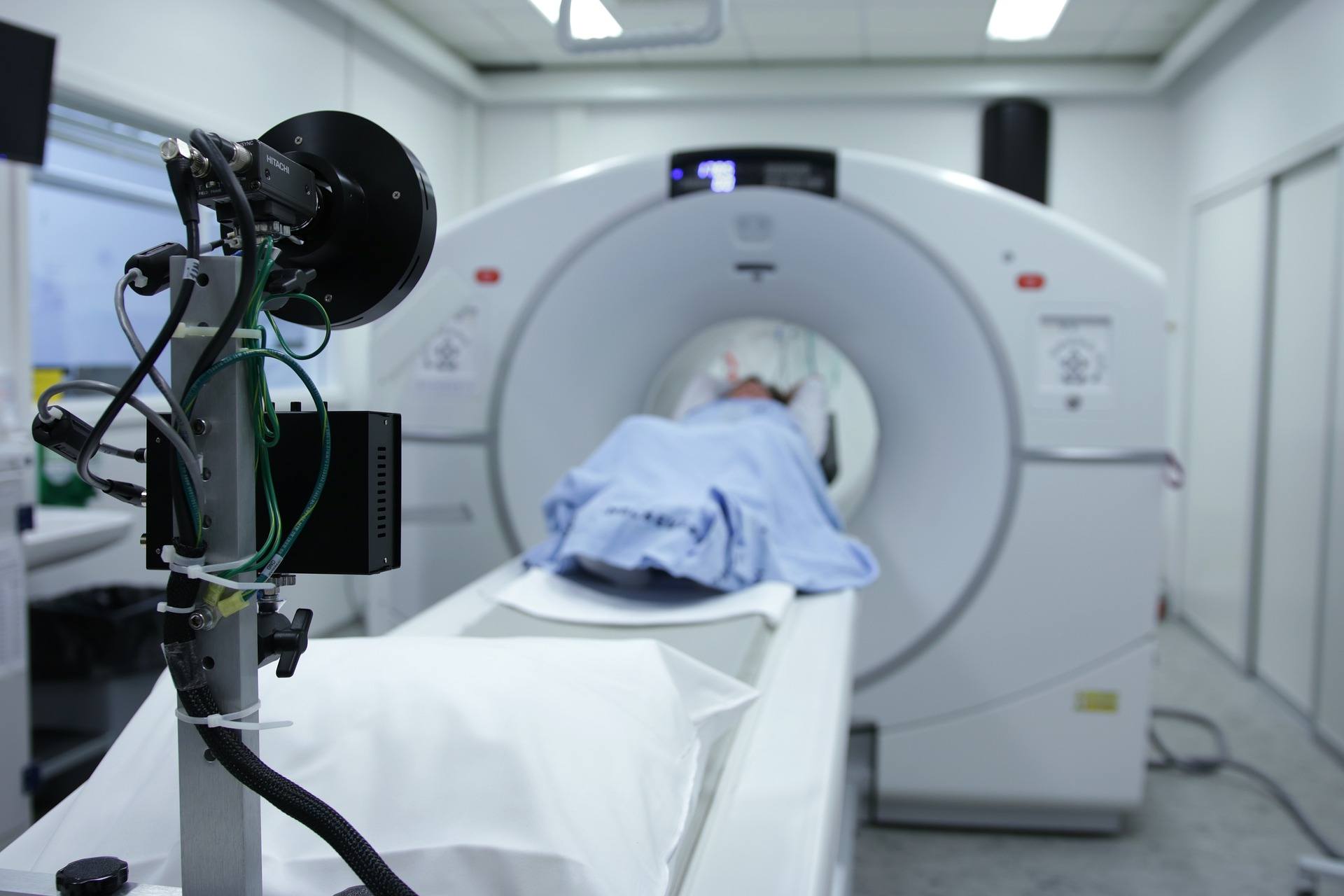
Mesothelioma is a rare and aggressive form of cancer that is often difficult to identify and accurately diagnose. Veterans who are currently undergoing screening for pleural mesothelioma diagnosis should be aware of the 4 main types of imaging tests and the difference between them.
Diagnosing pleural mesothelioma is complicated because it shares symptoms with other diseases that are more common. To accurately diagnose pleural mesothelioma, doctors conduct various imaging tests and perform a final tumor biopsy to confirm the diagnosis.
Veterans undergoing mesothelioma testing should understand the different tests and feel comfortable making informed decisions regarding their treatment and care.
Types of Mesothelioma Imaging Tests
Many doctors are not familiar with asbestos-related diseases. Pleural mesothelioma is often mistaken as lung cancer or a less serious illness. Because pleural mesothelioma is commonly misdiagnosed at first, doctors now use many imaging tests to accurately identify and stage mesothelioma.
Using imaging tests to diagnose cancer is a field of medicine called diagnostic radiology. Doctors use these tests not only to confirm there is a tumor mass present but also to know exactly where the tumor is located and how far the cancer has spread.
The 4 main types of imaging tests in pleural mesothelioma diagnosis are:
- X-rays
- Computerized tomography (CT) scans
- Positron emission tomography (PET) scans
- Magnetic resonance imaging (MRI) tests
X-Rays
X-rays generally help doctors find cancer in bones and organs. In the case of pleural mesothelioma, x-rays help doctors find tumors in the lung lining. X-rays are fast, painless and don’t require special preparation.
A special tube inside the x-ray machine delivers a beam of radiation to the body. Tissues within the body absorb or block the radiation to varying degrees and produce a 2-dimensional image. Bones show up as black, soft tissues as gray and organs that are mostly air (lungs) normally look black.
Tumors are generally denser than surrounding tissues and, therefore, show up as light shades of gray.
X-rays expose the body to radiation, but modern equipment uses much smaller amounts of radiation than in previous years. X-rays are good at finding bone problems, but CT scans and MRIs often give better pictures of organs and soft tissues.
CT Scans
Computerized tomography (CT) scans can help doctors find tumors and show details such as the tumor’s shape and size. The imaging test is painless and generally takes between 10 and 30 minutes to complete.
CT scans show bones, organs and soft tissues more clearly than standard x-rays. CT scans use a thin beam to create a series of images taken from different angles. The collected data is put into a computer, which creates a black and white image of a particular cross-section in the body.
If you were to imagine your body as a loaf of bread, looking at a CT scan would be like looking at a single slice.
When looking at a CT scan, doctors can identify a tumor, learn about its size and location. They also learn more about any surrounding blood cells that are feeding the tumor and causing it to grow. By comparing CT scans over time, doctors can see if a patient is responding to treatment or confirm if the mesothelioma has come back (recurrence).
PET Scans
Positron emission tomography (PET) scans are a type of nuclear medicine scan that helps doctors stage a patient’s cancer—meaning the test shows how far the cancer has spread.
PET scans are painless and use a special dye with radioactive tracers, also known as a contrasting agent, to identify diseased locations in the body. Once swallowed, inhaled or injected, this contrast agent collects in areas of higher chemical activity, which generally indicates the presence of disease.
The high chemical activity areas that indicate cancer show up as bright spots on PET scans.
PET scans show a wide range of activity on a cellular level and should be carefully reviewed by your doctor. It is possible for PET scans to identify noncancerous conditions and fail to identify solid tumors.
MRI
Magnetic resonance imaging (MRI) tests help doctors find cancer and look for signs that the cancer has spread. MRI is painless and requires no special preparation, but it is important to tell your medical professional if you have any metal in your body, just as from a medical implant.
Unlike x-rays and CT scans, MRI uses strong magnets to make images instead of radiation. MRI produces sliced images of the body from various angles that show pictures of soft tissues in the body that are often hard to see using other imaging tests.
Apart from diagnosis, MRI images can also help doctors plan treatment interventions such as surgery and radiation.
Pleural Mesothelioma Diagnosis in Veterans
Imaging tests are the first step in achieving an accurate pleural mesothelioma diagnosis. It is important for vets to know that the only conclusive way to diagnose mesothelioma from other common forms of illness is through the collection and testing of a tissue sample (biopsy).
Tissue samples will confirm the type of cancer and specific mesothelioma cell type. The earlier pleural mesothelioma can be detected, the more treatment options veterans have, increasing the chance of survival. If you are a veteran and have any hesitations or concerns regarding your recent mesothelioma diagnosis, it is important you seek a second opinion.
Contact the Mesothelioma Veterans Center today to learn more about your eligibility for VA benefits and gaining access to top mesothelioma specialists and treatments near you.

